 It’s always my steadfast mission to know which surgical methods will make patients feel more at ease and reassured that their health is in the best hands possible. This is certainly the case when a biopsy or lumpectomy is required.
The standard procedure when women need to have a cancerous mass removed, or a lumpectomy, is to place a needle in the breast to direct the surgeon to the tumor. This is called needle localization, and it’s done before a lumpectomy to help identify the precise location of a mass or tumor that cannot be palpably felt. Until surgery occurs, the needle is left in place or, more typically, a wire is inserted in its place.
While preoperative needle localization has been the standard procedure for a biopsy or lumpectomy of nonpalpable breast cancer tumors, it has drawbacks. The biggest disadvantages of needle localization are patient discomfort and rates of missing the target, i.e. needing to reposition the needle if it’s placed incorrectly, or if it migrates or drifts after being placed.
A technique I use called Hematoma Ultrasound-Guided (HUG) bypasses the need for needle localization for breast cancer lumpectomies, and this procedure offers several advantages over needle localization.
HUG Is More Comfortable, Accurate, Faster & Easier
In most cases, the diagnosis of early-stage breast cancers are confirmed by an image-guided core needle biopsy. The core breast biopsy procedure usually creates a small cavity at the site of the tumor which turns into a hematoma, meaning a blood ball resulting from the initial core needle biopsy.
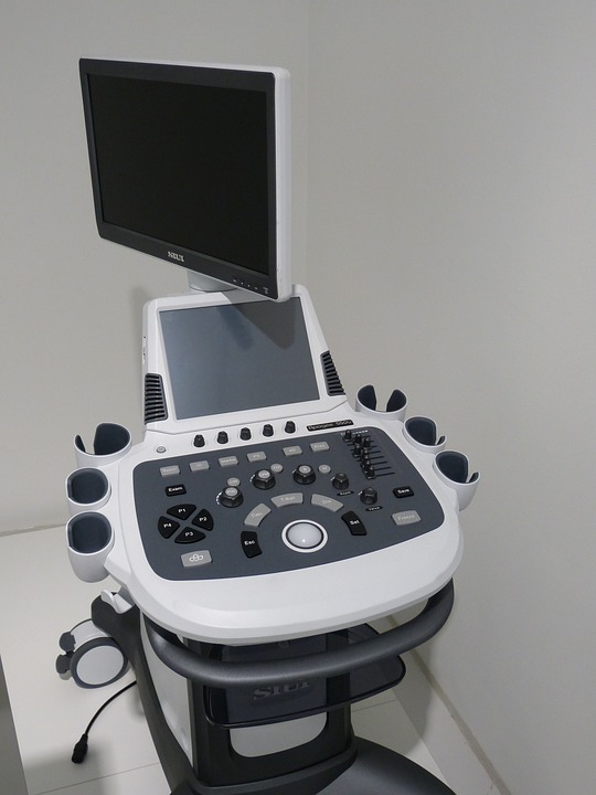
In the early 2000’s, researchers began to test the theory that if there is a hematoma, then a needle localization procedure may not be needed. This is because the hematoma can be seen on ultrasound, so a surgeon can accurately find and excise the actual biopsy site of non-palpable breast lesions without having to rely on a needle or wire to show the location.
If a hematoma has not already formed after a core needle biopsy, then one can be placed several days before the surgery by injecting the patient’s own blood into the breast to target the non-palpable lesion. This makes scheduling easier and also eliminates the risk of migration that may occur with needle localization.
This new procedure is often more comfortable for the patient because no wire or needle is left in the breast. It’s technically faster and easier because ultrasound is used to directly show the location of the hematoma at surgery and to confirm lesion removal in the operating room by specimen ultrasound. Ultimately, by eliminating the additional procedure needed for needle localization, HUG can be both more time and cost-efficient for the patient and surgeon.
In addition, research studies reported in the National Institute of Health’s Center for Biotechnology Information suggest that HUG is more accurate in localizing and removing non-palpable lesions than needle localization.
I continue to be encouraged about advances in improved imaging methods for the detection and treatment of breast cancer, and find that HUG, when applicable, is preferable for patient comfort and surgical accuracy.
If you’ve recently been diagnosed with breast cancer or are wondering if you should seek a second opinion on your diagnosis or surgical plan, I’m here to consult with you.
Committed to serving breast cancer patients through my solo practice in Breast Surgical Oncology and General Surgery, I have offices at Tennova Turkey Creek Medical Center in West Knoxville, at Tennova North Knoxville Medical Center in Powell, at Jefferson Memorial Hospital, and in Newport. My extensive research and dedication to continual learning have distinguished me as a leader in the field of specialized breast cancer oncology. To learn more about my compassionate surgical care approach, visit www.aaronmd.com or call (865) 692-1610.